


Achilles Tendon
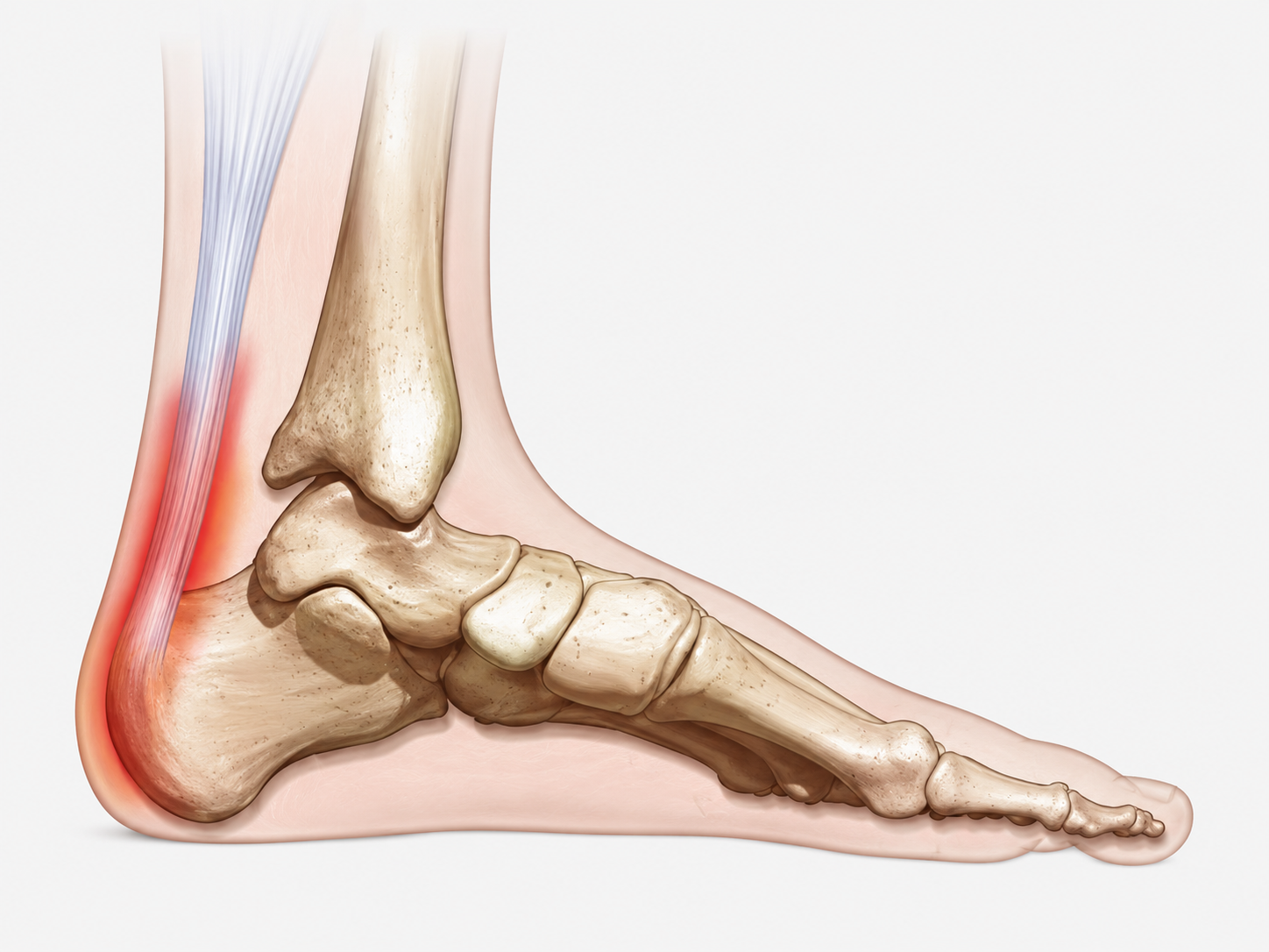
Achilles tendinopathy may involve the tendon above the heel or the insertion where the tendon attaches to the calcaneus. Tendon load, calf function, training volume and compression around the insertion can all influence symptoms.

Pain behind the heel can arise from the Achilles tendon, the bursae around the tendon, the posterior heel bone, or a combination of structures. Identifying which structure is involved changes what should happen next.
Posterior heel pain often follows a recognisable pattern. Patients commonly report one or more of the following.
Pain on the first few steps after rest
Stiffness after sitting or periods of inactivity
Discomfort during push-off, hills or stairs
Localised tenderness or swelling behind the heel
Symptoms that settle, then return with activity
Patients with pain in the back of the heel have often already tried rest, stretching, massage, calf raises, footwear changes or another form of treatment.
Sometimes symptoms improve temporarily. Sometimes they do not change at all.
One reason is that posterior heel pain is often treated as though every case is the same. It is not. The Achilles tendon, surrounding bursae and posterior calcaneus have different tissue properties and different tolerances to load and compression.
A treatment that is appropriate for one structure may be poorly timed or inappropriate for another.
The location of pain may be similar, but the structure responsible can be different.
Achilles tendinopathy may involve the tendon above the heel or the insertion where the tendon attaches to the calcaneus. Tendon load, calf function, training volume and compression around the insertion can all influence symptoms.
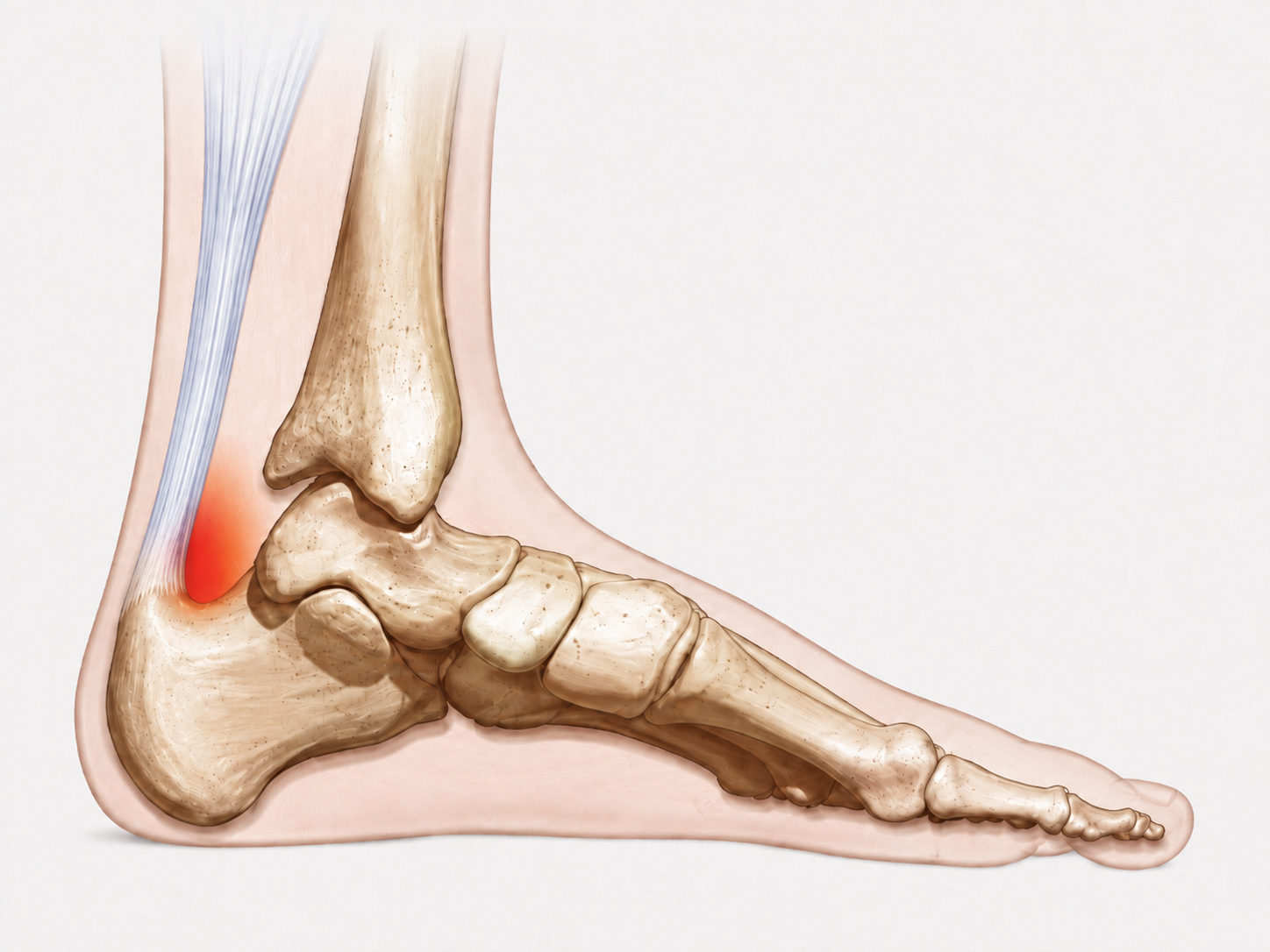
Retrocalcaneal bursitis and irritation of the superficial bursa can produce tenderness, swelling and pain around the posterior heel, particularly where local compression is present.
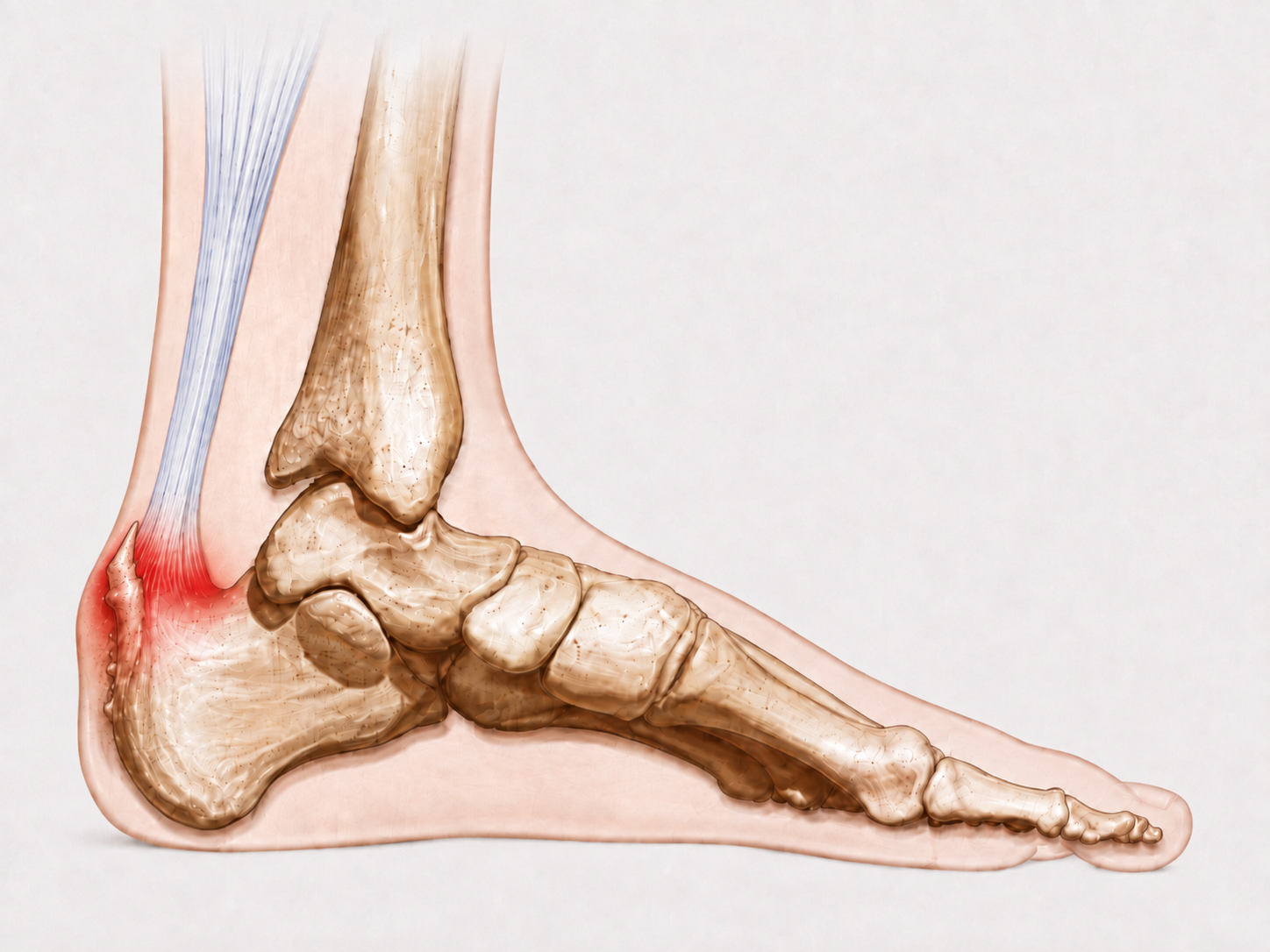
A prominent posterior calcaneus may increase local pressure around the Achilles insertion and adjacent soft tissues. Footwear shape, heel-counter pressure and repetitive compression can all influence symptoms.
Clinical investigation should establish the diagnosis, identify the mechanical drivers behind the problem, and determine which clinical and biomechanical findings are relevant to the individual patient.
Correctly identifying the symptomatic tissue is fundamental. Failure to diagnose the structure responsible can direct subsequent treatment towards the wrong tissue from the outset.
Clinical history and examination are used to diagnose the tissue or structures responsible for the symptoms and differentiate between other potential causes of posterior heel pain.
Mechanical loading patterns, occupational demands, habitual foot positions, training exposure, calf function, footwear interaction and repetitive compressive or tensile forces are evaluated to identify the factors perpetuating tissue stress.
Gait, foot and ankle function, joint movement, calf function and footwear are assessed to determine which biomechanical findings are clinically relevant to the individual presentation.
Clinically identify which load modifications are required, which mechanical driving forces need to be reduced or removed, and determine the appropriate sequence in which treatment interventions should then be introduced.
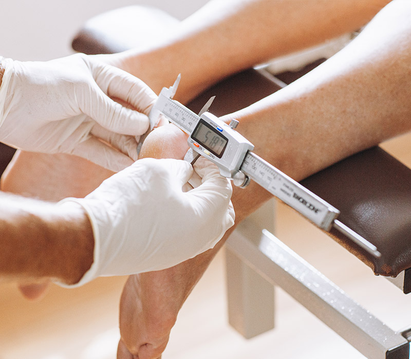
Once the diagnosis and relevant mechanical drivers have been established, treatment can be selected and sequenced according to the clinical findings rather than applying the same intervention to every presentation.
The treatment itself is only part of the equation. Selecting the correct intervention, at the correct stage, for the correct tissue is what matters.
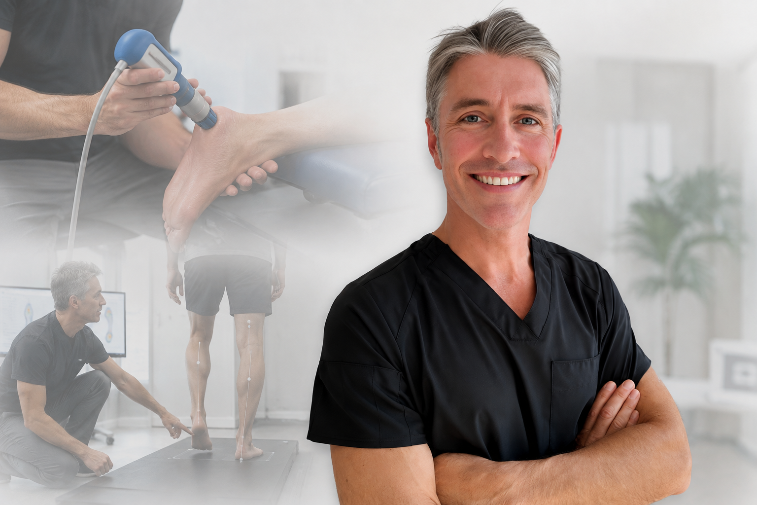
Sydney Heel Pain Clinic is focused on the investigation and treatment of heel and lower-limb pathology, combining clinical diagnosis, biomechanical assessment and treatment planning within the same clinical process.
The consultation begins with your history, followed by clinical examination and investigation of the factors contributing to the problem.
We listen first. Your backstory matters. When did the pain begin? What were you doing at the time? What treatments have you already tried? What aggravates it, what settles it, and how has it changed over time?
Examination is used to diagnose the symptomatic tissue and differentiate between the possible causes of pain in the posterior heel.
Gait, foot mechanics, calf function, footwear, activity and relevant loading patterns are investigated to identify the contributing mechanical drivers.
The diagnosis and contributing factors are explained, required load modifications are identified, and the appropriate treatment strategy is established.
You leave with an understanding of the diagnosis, the factors contributing to it and the rationale behind the treatment plan.
Kim Brierley
Google review
“Fantastic clinic. I had Achilles tendonitis which was extremely painful. I had seen another podiatrist who gave me exercises that made my condition worse. The shock treatment and new orthotics have worked wonders. I highly recommend this clinic.”
Bao Pham
Google review
“My heel pain problem had been going on for over 6 years. I've been through several physio therapy clinic, the issue only got slightly better but never completely heal. After two sessions with Karl, for the first time in 6 years, I didn't feel pain in the morning and didn't need to tip toe for a few minutes after getting out of bed.”
Emmanuel Giannaros
Google review
“For almost 4 years I was barely able to walk for 2–3 days after participating in any high impact sport/activity. After just one consultation with Karl, he provided me with injury specific educational information, recommendations on appropriate footwear and he also designed custom made orthotics for me. For the first time in 4 years, I was able to return to playing competitive sport.”
Read further patient experiences on Google.
If posterior heel pain is persisting, repeatedly returning, or changing the way you walk, exercise or choose footwear, establishing a diagnosis becomes more useful than continuing to trial different treatments without knowing which tissue is involved.
The Achilles tendon transfers force from the calf muscles to the heel during walking, running and push-off. Pain may occur within the tendon above the heel or at the insertion where the tendon attaches to the calcaneus.
In insertional Achilles tendinopathy, both tensile load and compression around the posterior heel may be relevant. Symptoms can therefore be influenced by training load, calf function, ankle movement, footwear and the shape of the posterior calcaneus.
The retrocalcaneal bursa sits between the Achilles tendon and the posterior calcaneus. Irritation in this region may produce pain, local tenderness and swelling.
Compression from footwear, repetitive loading and nearby structural prominence can contribute. Distinguishing bursitis from Achilles tendon pain is clinically important because management may differ.
Haglund's deformity describes a prominent area at the posterosuperior calcaneus. In some patients this prominence increases compression against the Achilles insertion, surrounding bursae or footwear.
The presence of a bony prominence does not automatically establish it as the sole cause of symptoms. Clinical findings need to be considered together with footwear pressure, local tissue irritation and mechanical load.
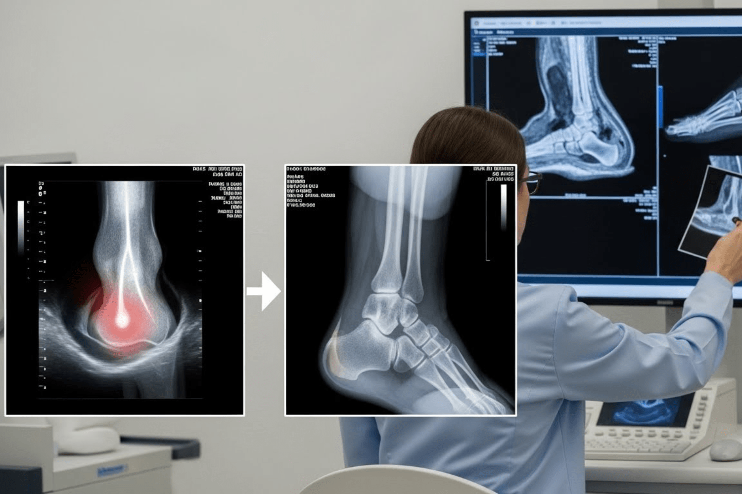
Imaging is not required in every presentation of posterior heel pain. Where the clinical presentation or differential diagnosis warrants further investigation, X-ray, ultrasound or other imaging may be considered to assess bone, tendon and surrounding soft tissues.
Treatment is directed by the diagnosis and the mechanical factors contributing to continued tissue load or compression.
Depending on the clinical findings, management may incorporate load modification, calf rehabilitation or stretching programmes, footwear-specific recommendations, current-market footwear advice, shockwave therapy, heel lifts and 3D-printed custom footbeds.
The sequence of treatment can be as important as the intervention itself, particularly where tissue irritability, local compression or suspected tendon tearing needs to be considered.
Aldridge T. Diagnosing heel pain in adults. American Family Physician. 2004;70:332–338.
Hendrix CL. Calcaneal apophysitis (Sever disease). Clinics in Podiatric Medicine and Surgery. 2005;22:55–62.
Pearce CJ, Tan A. Non-insertional Achilles tendinopathy. EFORT Open Reviews. 2016;1:383–390.
Vaishya R, Agarwal AK, Azizi AT, Vijay V. Haglund's Syndrome: A Commonly Seen Mysterious Condition. Cureus. 2016;8(10).