Case study 27 June, 2016 – Sever’s Disease
A 28-year-old male presents to the clinic complaining of excruciating pain at the back of his right leg – near the Achilles tendon area. He reports that the pain came on after a session of playing soccer 3 days ago, after having a 4-month break. He has been limping on the right foot since, and experiences frequent throbbing at the sore region. He has been advised to use NSAIDs by his local doctor, which have been helpful in reducing pain temporarily.
Upon visual examination, the Achilles tendon is swollen and red about 6 cm from its insertion on to the back of the heel bone, as is commonly seen in Achilles Tendonitis. On physical examination, palpation on the middle of the tendon elicits significant pain. No bulbous mass on the tendon or its insertion is noted – which would otherwise indicate a more chronic pathology. The patient is informed that he has Achilles Tendonitis.
A thorough biomechanical assessment is conducted, whereby the patient is asked to walk and run on a treadmill, as his walking and running form is analysed and video recorded. It is noted that he is a heavy heel striker, and pronates mildly through his ankle and sub talar joints. Furthermore, he has tightness in his calf muscles and therefore has a limited range of ankle joint dorsiflexion.
All of the above increase the pull through the Achilles tendon and exacerbate his condition by adding strain.
A footwear assessment reveals that the back soles in his running shoes are worn out and compressed significantly.
It is explained to the patient that he has an acute onset of aggressive Achilles Tendonitis – which is inflammation of the tendon resulting from micro-tears caused by overloading the tendon with a tensile force that is too heavy and/or too sudden. An Ultrasound report confirms that there are no tears.
TREATMENT
Due to the severity of the inflammation, the focus of treatment is to unload and completely immobilise the Achilles tendon to initiate healing. This is done by fitting an immobilisation boot to his right leg. He will wear the boot until his symptoms decrease or resolve, usually within 4-8 weeks. A heel lift is also added inside the boot to elevate the heel and reduce tension on the Achilles tendon.
He is also advised to ice the area and stretch his calf muscles every night, with the correct stretch technique demonstrated.
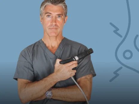
The injured tendon is also treated with the Extracorporeal Shockwave Therapy machine to increase blood flow and stimulate tissue regeneration, aiding in faster recovery. This is done at weekly intervals for 5 weeks. Shock wave therapy is a very reliable treatment option for Achilles Tendonitis and other conditions that require blood flow such as Plantar Fasciitis.
Achilles Tendonitis – Follow up
Pain completely subsided within the estimated 6-8-week period. In the long term, he was advised to change his running form, as well as incorporating more frequent calf stretching exercises.
Please note that there are several treatment options for heel pain and similar conditions such as Plantar Fasciitis, Achilles Tendonitis / Tendinosis, Sever’s disease and Bursitis. The most appropriate treatment option for each individual patient can only be selected after a physical examiniation and biomechanical assessment has been carried out.
For more information on other conditions such as Plantar Fasciitis please visit www.sydneyheelpain.com.au/what-is-plantar-fasciitis/
Written by Karl Lockett


